Advanced diffusion weighting: double wave vector diffusion weighting
Diffusion-weighted nuclear magnetic resonance has proven very successful in obtaining information about the microscopic structure of biological tissues. Pathological processes can alter the diffusion characteristics of water in tissue significantly. The most common method of diffusion weighting is based on a pair of two magnetic field gradient pulses. This project is based on two such pairs with different gradient direction. Theoretical and experimental pioneering work predicts that the measured signal can depend on the angle between these directions if diffusion is restricted by the boundaries of cells or compartments. The details of this angular dependence are determined by the size and shape of the compartments.
The project sets out to develop new non-invasive methods for measuring cell size and shape in vivo on the basis of this phenomenon. Such techniques are anticipated to extend the existing methods for investigation and diagnosis of pathologically altered tissues, such as in cancer or degenerative diseases of the human brain. The results will be compared with other measures for characterizing tissue structure, such as diffusion kurtosis, e.g. It is to be shown whether the method supplies additional information or outperforms existing methods.
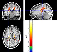
The colour-code in the figure represents a size estimate based on double wave vector diffusion weighting in a region of interest in the corticospinal tracts of a healthy volunteer.
Grants
- German Research Foundation under grant number DFG KO3389/2
- previous: German Research Foundation under grant number DFG KO3389/1
Former Project Members
- Vincent Méthot
- Viktor Wottschel
- Lars Kreutzburg
- Patricia Ulloa
- Tatiana Nemţanu
Cooperations
- Universitätsklinikum Schleswig-Holstein
- Universitätsklinikum Hamburg-Eppendorf
- Philips Healthcare, Hamburg
- Siemens Healthineers
Publications
-
Extra-axonal contribution to double diffusion encoding-based pore size estimates in the corticospinal tract, Magnetic Resonance Materials in Physics, Biology and Medicine, 36(4), 589–612, 2023, DOI: 10.1007/s10334-022-01058-8.
-
Bias in the apparent exchange rate measurements: insight from numerical simulations, 1085, 2017.
-
Pore size estimation from double diffusion encoding, Current Directions in Biomedical Engineering, 30(2), 627–630, 2017, DOI: 10.1515/cdbme-2017-0131.
-
Experimental validation of a bias in apparent exchange rate measurements, Current Directions in Biomedical Engineering, 3(2), 529–532, 2017, DOI: 10.1515/cdbme-2017-0112.
-
Varying the mixing time of the double diffusion experiment: A better experimental design for pore size estimation, Magnetic Resonance Materials in Physics, Biology and Medicine, 30 (Suppl. 1), S633-S634, 2017, DOI: 10.1007/s10334-017-0635-y.
-
Pore size estimation using the mixing time dependence of a double diffusion encoding experiment: experimental validation on a clinical MR system, 717, 2017.
-
Experimental study of bias in apparent exchange rate measurements, S173-S174, 2017, DOI: 10.1007/s10334-017-0633-0.
-
Pore size distribution estimation using the mixing time dependency of a double diffusion encoding experiment: a proof of concept from Monte Carlo simulated data, S165-S167, 2016, DOI: 10.1007/s10334-016-0569-9.
-
Exploring model limitations for double diffusion encoding MRI, 2016.
- [ BibTeX ]
-
Determination of the pore size distribution by double diffusion encoding, 12, 2016, DOI: 10.3389/conf.FPHY.2016.01.00012.
-
Conventions and Nomenclature for Double Diffusion Encoding NMR and MRI, Magnetic Resonance in Medicine, 75(1), 82–87, 2016, DOI: 10.1002/mrm.25901.
-
Design of a Motion Phantom for Magnetic Resonance Imaging, 251–254, 2016.
- [ BibTeX ]
-
Water-in-oil Emulsion as a Simple Phantom for Validation of Double Diffusion Encoding MRI Sequences, S48-S49, 2016, DOI: 10.1007/s10334-016-0568-x.
-
Studying the extracellular contribution to the double wave vector diffusion-weighted signal, Current Directions in Biomedical Engineering, 1(1), 240–244, 2015, DOI: 10.1515/cdbme-2015-0060.
-
Investigating the Extracellular Contribution to the Double-Wave-Vector Diffusion-Weighted Signal, 2786, 2015.
- [ BibTeX ]
-
Extracellular Space Contribution to the Double Wave Vector Diffusion-Weighted Signal, s568, 2014, DOI: 10.1515/bmt-2014-5008.
-
Chasing the Zebra. The Quest for the Origin of a Stripe Artifact in Diffusion-Weighted MRI, s535-s538, 2014, DOI: 10.1515/bmt-2014-5008.
-
Visualizing Microscopic Hemorrhages with Susceptibility-Weighted Imaging (SWI) for Forensic Applications, s539-s542, 2014, DOI: 10.1515/bmt-2014-5008.
-
Double Wave Vector Diffusion Weighting in Wallerian Degeneration, 3564, 2012.
- [ BibTeX ]
-
Towards compartment size estimation in vivo based on double wave vector diffusion weighting, NMR in Biomedicine, 24(10), 1422–1432, 2011, DOI: 10.1002/nbm.1711.
